Monday, November 28, 2011

Answer to the last post: it was a unicorn...

Well, not really... at least not the horse-type of unicorn, those can only exist in the imagination of people who want to be marine biologist because they think that by doing so, they will get to train and play with dolphins at some theme park...
Anyways, the picture on the previous post seems to have been quite straightforward after all. Yes, it is part of a narwhal tusk, so congrats to all who guessed right! Narwhals, which go by the scientific name Monodon monoceros, are odontocetes (toothed whales), distinguished by the long spiraled tusk seen in the males (and occasionally females) of the species, and are thus sometimes called the "unicorn of the sea".
Dorsal view of a double-tusked narwhal, on exhibit at the Sant Ocean Hall at the National Museum of Natural History in Washington, DC. Normally, narwhals only have one enlarged tusk (the left one) so this one is an exception.
The closest relatives of the narwhals are the belugas or white whales (Delphinapterus leucas); together these two species comprise the family known as Monodontidae. Among extant toothed whale groups, monodontids are more closely related to porpoises (Phocoenidae) and to dolphins (Delphinidae) (e.g. see Geisler et al., 2011), whereas relationships with extinct groups include Albireonidae (Barnes, 1984) and Odobenocetopsidae (de Muizon, 1993). Odobenocetops previously featured here, also has an enlarged tusk-like tooth like Monodon. However, the tusks are located in different bones within the rostrum (the premaxilla in Odobenocetops, the maxilla in Monodon [see below]), and hence although somewhat similar, they are probably not homologous.
Ventral view of the rostrum of extant monodontids.
The fossil record of monodontids, is scarce (at least when compared to that of other living odontocete groups). The oldest monodontid known is Denebola brachycephala from the late Miocene of Baja California (Barnes, 1984). Other fossil monodontids are known from early Pliocene deposits in the North Sea (Lambert and Gigase, 2007) and the eastern coast of North America (Withmore, 1994; Withmore and Kaltenbach, 2008; Kazár and Bohaska, 2008)*. All these fossil forms are more like Delphinapterus, i.e. they have multiple teeth in the maxilla, whereas Monodon only has an enlarged tusk (usually only in the male) and the females is edentulous (see illustration above). So how did the tusk of narwhals came to be or what is it for? It is still a bit of a mystery!
*I'll blog more on these North American ones sometime next year, so stay tuned!

For a neat article on the tusk and on monodontids visit this page over at Tetrapod Zoology v2.
And here if you or someone you know wants to be a marine biologist.

References:

Barnes, L. G. 1984. Fossil odontocetes (Mammalia: Cetacea) from the Almejas Formation, Isla Cedros, Mexico. PaleoBios 42:1–46.

Geisler, J. H., M. R. McGowan, G. Yang and J. Gatesy. 2011. A supermatrix analysis of genomic, morphological, and paleontological data from crown Cetacea. BMC Evolutionary Biology 11:112.

Kazár, E., and D. J. Bohaska. 2008. Toothed whale (Mammalia: Cetacea: Odontoceti) limb bones of the Lee Creek Mine, North Carolina; pp. 271-324 in C. E. Ray, D. J. Bohaska, I. A. Koretsky, L. W. Ward, and L. G. Barnes (eds.), Geology and Paleontology of the Lee Creek Mine, North Carolina, IV. Virginia Museum of Natural History Special Publication 14.

Lambert, O., and P. Gigase. 2007. A monodontid cetacean from the Early Pliocene of the North Sea. Bulletin de l’Institut Royal des Sciences Naturelles de Belgique, Sciences de la Terre 77:197–210.


Muizon, C. de. 1993. Walrus-like feeding adaptation in a new cetacean from the Pliocene of Peru. Nature 365:745–748.


Whitmore, F. C., Jr. 1994. Neogene climatic change and the emergence of the modern whale fauna of the North Atlantic Ocean. Proceedings of the San Diego Society of Natural History 29:223–227.

Whitmore, F. C., Jr., and J. A. Kaltenbach. 2008. Neogene Cetacea of the Lee Creek Phosphate Mine, North Carolina; pp. 181–269 in C. E. Ray, D. J. Bohaska, I. A. Koretsky, L. W. Ward, and L. G. Barnes (eds.), Geology and Paleontology of the Lee Creek Mine, North Carolina, IV. Virginia Museum of Natural History Special Publication 14.

Monday, November 7, 2011

What is this?

This might be an easy guess for some of you or maybe not... Let me know what you think in the comments section. I'll be back with the answer in a week or so. Enjoy!

Saturday, September 3, 2011

Bad weather, ancient oceans and modern seagrasses… a brief visit to Puerto Rico


Summertime is normally when geologist and paleontologist engage in fieldwork. For me the last two summers have been different; I’ve spent both teaching in the human anatomy lab at USUHS, which has been a fantastic experience. However, being busy in the summer months hasn’t really stopped me from doing a little fieldwork, fortunately, I work mostly in the Caribbean region where the weather is generally nice and the only thing to worry about is hurricane season…

And so it was that I had the chance to go to Puerto Rico at the end of the summer and in the midst of hurricane season. Every time I go there is both for pleasure and work; I get to see my family, enjoy the food and scenery and with a healthy dose of fieldwork, it makes for the perfect vacation. This time I arrived two days before hurricane Irene (then still a tropical storm) hit the island. I was fortunate to be staying in the northwestern part of the island, which did much better than most other parts. We got rain, lots and lots of it, which kind of messed up my plans for fieldwork a little bit.

Meanwhile, while it rained, I stayed busy looking through fossils I had collected during my years as an undergrad; mostly fish teeth (bony and cartilaginous) and otoliths, which I’ll use for some projects on the works. It wasn’t until five six, days after my arrival that it was sunny and dry enough to go out to the field. Without much time to go to different places, I decided that my best chances for fieldwork were in the vicinity of my hometown, basically here and here.

The early Oligocene "mystery" bone I found.

So first I went to the Río G locality where early Oligocene deposits are exposed along the banks of the river. Its one of my favorites and almost always I find something of interest. With all the rain, I was both worried and hopeful. Worried that cool stuff was being eroded away, but hopeful in that new stuff would be exposed. After prospecting for a while and not finding anything more than some sirenian ribs, I made it to the “bonebed” (previously featured here). This time around all that I found was an interesting piece of bone (see picture above), and as it turned out, I had good reasons be worried. If you look at the picture, the part of the bone towards the bottom had broken off, recently, probably during the rain and increased water levels earlier in the week. It doesn’t seem to be a rib, which is why I think its interesting, but due to its incompleteness I’m still not sure what it is… it sounds a bit frustrating, but the best thing to do is to turn that frustration into motivation to keep looking!

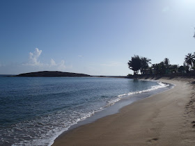
 My favorite beach in the Atlantic coast of Puerto Rico. Just how I like it, early in the morning and nearly devoid of people.

My other field excursion was to the beach. Working with marine organisms that thrive in shallow marine environments is always a good excuse for a trip to the beach (specially in the tropics). Staying in my hometown meant that my favorite beach on the Atlantic coast of Puerto Rico was only a 15-minute drive away! I didn’t see any manatee (wasn’t expecting to anyways) but did get to see some seagrass beds (see picture below).

It was wavy and shallow (~1m) hence all the suspended sediment. The turtle grass blades (leaves) are about 0.5-1 cm wide and ~10-12 cm long.

Most seagrass beds in the Western Atlantic and Caribbean region consist of several species, with Thalassia testudinum (turtle grass), Halodule beaudettei (shoal grass), and Syringodium filiforme (manatee grass) as the more common ones. However, the seagrass bed that I was looking at was monospecific (= only one species present). As far as I could tell it consisted solely of turtle grass. Monospecific beds of turtle grass are not uncommon, in fact, Thalassia is what is known as a climax species. That means that it is the dominant species of seagrass; other species (like the ones mentioned above) are only present in small, disturbed patches or at the periphery of the beds. The dominance of turtle grass over other seagrass species and its consequences (like mass die-offs) in historical times has been partly blamed on overkill of large marine herbivores, mainly seacows and green turtles (Jackson et al., 2001). However, this could be slightly different when viewed from a deeper historical perspective. I hope to bring more on this sometime in the upcoming months…


Jackson, J. B. C. et al. 2001 Historical overfishing and the recent collapse of coastal
ecosystem. Science 293, 629-638.

Wednesday, June 15, 2011

The hand of Steller’s sea cow, revisited

It’s been quite a while since my last post. A lot has been going on, mostly research-related, which is good. Just last week I was in San Diego where I participated in the Sixth Triennial Conference of Secondary Adaptation of Tetrapods to Life in Water held at San Diego State University. The meeting was a great opportunity to see colleagues as well as making new acquaintances, hat tip to the host committee: Annalisa Berta, Tom Deméré and Eric Ekdale for such a great meeting! The week before that I spent some days visiting the collection at the Natural History Museum of Los Angeles where I got to look at some interesting sirenian and cetaceans, many thanks to Larry Barnes and Sam McLeod for access to specimens.

Ok, so now to the meaty part of this post. Back in 2008 I wrote about the hand of Steller’s sea cow (Hydrodamalis gigas) or more likely the lack of one. Back then I mentioned that G. W. Steller was one of the few people who saw H. gigas alive, and in his account he mentions the lack of fingers in this species. However, so far no hand bones have been found associated with Hydrodamalis gigas or even H. cuestae (see reconstruction below) (the presumed direct ancestor of the former), this lead to skepticism about his observations. So, how was that Steller’s account about the hand of H. gigas was corroborated?

Hydrodamalis cuestae, in left lateral view (picture of the reconstruction on exhibit at the San Diego Natural History Museum). Notice the reduced forelimbs.

The discovery of Dusisiren dewana in the Late Miocene (10-8 million years ago [mya]) of Japan, provided the key elements to support Steller’s account. Dusisiren is a genus of sea cow that include two other, earlier species: D. reinharti from the Early Miocene (20-16 mya) and D. jordani from the Middle-Late Miocene (12-10 mya) (see picture of D. jordani below). The species of Dusisiren together with Hydrodamalis cuestae (see picture above), H. spissa and H. gigas are members of a subfamily of dugongids (which means they are more closely related to dugongs than to manatees) known as the Hydrodamalinae (Domning, 1994; Domning & Furusawa, 1994). Hydrodamalines, as they are also known, ranged throughout coastal waters of the northern Pacific, from Baja California to Japan, with one species surviving until historical times (Domning & Furusawa, 1994). Some distinctive features of this group include, large body size, reduction or loss of dentition, and reduction of the digits, which brings us back to D. dewana.

Dusisiren jordani, anterior view (specimen at exhibit at the Natural History Museum of Los Angeles). Notice the "normal" hand in this species.

Described by Takahashi et al. in 1986, the remains of Dusisiren dewana were found in Late Miocene deposits in Japan and together with other elements of the skeleton there was also a nearly complete forelimb, including the hand. These were crucial in providing an insight into what the hand of later hydrodamalines was like. Dusisiren dewana shows reduction of the hand bones, in this respect differing from D. jordani, its predecessor, whose hands showed no reduction of elements (i.e. long metacarpals and long fingers composed of multiple elements) (see picture above). As for D. dewana, the carpals (wrist bones) are quite similar to those of other sirenians, however, the metacarpals (the bones between the wrist and fingers) and the phalanges (finger bones) are reduced, actually extremely reduced in the case of the phalanges (see picture below of Hydrodamalis cuestae). D. dewana as the sister taxon (closest relative/ancestor) of Hydrodamalis spp. is best evidence at hand (unintended pun) supporting Steller’s account on the hand of the sea cow that bears his name and allowing for reconstructions like the one seen here for H. cuestae.

Hydrodamalis cuestae, right lateral view (picture of specimen on exhibit at the San Diego Natural History Museum). Notice the reduced hand.

Domning, D. P. 1994. A phylogenetic analysis of the Sirenia. Proceedings of the San Diego Society of Natural History 29:177-189.

Domning, D. P. & H. Furusawa. 1994. Summary of taxa and distribution of Sirenia in the North Pacific Ocean. Island Arc 3:506-512.

Takahashi, D., D. P. Domning & T. Saito. 1986. Dusisiren dewana, n. sp. (Mammalia: Sirenia), a new ancestor of Steller’s sea cow from the upper Miocene of Yamagata Prefecture, northeastern Japan. Transactions and Proceedings of the Paleontological Society of Japan, New Series 141:296-321.

Saturday, March 5, 2011

When you're strange, or how different are the teeth of extant sirenians

Living sirenians (sea cows: manatees and dugongs) are but the remnants of a much more diverse group whose history extends back at least to the late early Eocene (~50 million years ago [Ma]). There are only four living species of sirenians: the manatees (grouped in the family Trichechidae) and including three species: the Antillean manatee (Trichechus manatus)*, the Amazonian manatee (T. inunguis) and West African manatee (T. senegalensis). The other extant species is the dugong (Dugong dugon) that, together with the now extinct Steller’s sea cow (Hydrodamalis gigas), represents the family Dugongidae.

*T. manatus includes two subspecies: the West Indian manatee (T. manatus manatus) and the Florida manatee (T. manatus latirostris).

Above, Homo sapiens and Trichechus manatus latirostris. (Pic. courtesy of B. Bonde).

Although overall very similar to their extinct relatives, some characteristics of these extant sirenians are quite different and unique, not really representative of each particular group. One of those characteristics, and the one I will discuss here, is the dentition of Dugong dugon. This species, which lives in the Indo-Pacific region, is characterized by having molars that are best described as nearly enamel-less (a thin enamel cap is quickly worn off), open-rooted, ever-growing pegs (see pictures below), resembling more those of sloths, than those of manatees!!

Lower molars of Dugong dugon, notice that root and crown are continuous. The dark area is the part that was exposed above the gum line, hence the staining.

To understand how they differ let me give you a little background info on sirenian dentition. Primitively, the dental formula of sirenians consisted of three incisors, one canine, five premolars* and three molars (Domning, 1994; Savage et al., 1994). Throughout sirenian evolution, this formula has been reduced, first by loss of the fifth permanent premolar in the earliest dugongids (see figure below), then by subsequent loss of second and third incisors, canine and the other permanent premolars. In terms of general features, the molars of most fossil sirenians can be described as bilophodont, with enameled crowns that are clearly differentiated from the root (these being closed and multiple [with three in upper molars and two in the lowers]). These kind of molars somewhat resemble those of the most primitive proboscideans (Sanders et al., 2010), which, by the way, are the closest relatives of sirenians.

*Presence of a fifth premolar is reminiscent of the primitive eutherian condition and, among Cenozoic placental mammals, a pretty unique condition only seen in sirenians (Rose, 2006).

Partial left maxilla of a late Eocene (37.2-33.9 Ma) dugongid, (A) lateral view (reversed); (B) occlusal view (anterior to the right). Notice that the deciduous premolar is molarized and multi-rooted, whereas the permanent premolars are more conical and single rooted. dP = deciduous premolar; M = molar; P premolar (permanent).

Beginning about the early Oligocene (33.9-28.4 Ma) the dental formula of most dugongids (including Dugong) stayed the same; it consisted of the first upper incisor, upper and lower deciduous premolars 3-5 and permanent molars 1-3 (see A-B in figure below)* (Domning & Pervesler, 2001). Up until the appearance of Dugong dugon the morphology of the molars was, in general, much the same as that described above. The peculiar, somewhat aberrant, dentition of dugong likely evolved quite recently (geologically speaking). Unfortunately there are no fossils of D. dugon to really know when. The best evidence comes from a very Dugong-like sirenian skull from the Pleistocene of Florida (Domning, 2001) (sadly, it is in a private collection). This skull is from sediments that were deposited about two million years ago and is nearly identical to Dugong dugon; one of the few differences, actually the most obvious one, is that its teeth are much like those of earlier dugongids, bilophodont, with enamel and multiple, closed roots. Therefore, if this fossil is taken as the closest ancestor of extant dugong, it would imply that its bizarre dentition is a quite recent (less than 2 million years) evolutionary novelty.

*Or even further reduced from that condition (as was the case of Steller’s sea cow and its closest relatives).

Occusal view of various sirenians (A-C). A. Left upper toothrow of a late Oligocene (28.4-23 Ma) dugongid from Puerto Rico. B. Right lower toothrow of Dugong dugon, notice that the lack of enamel, leaves a nearly featureless surface; the black rim is just stain on the portion of the tooth that was above the gum line. C. Left upper toothrow of Trichechus inunguis.

Manatees (Trichechus spp.) also have unusual dentition when compared to dugongids and some of their ancestors (for starters they lack incisors). If you look at the figure above (C) you’ll notice that there seems to be more molars than there should be. And that is correct; manatees have supernumerary molars that are constantly being replaced throughout their entire life (not to be confused with the delayed eruption that characterizes elephants). The molars in Trichechus are also proportionately small, that way more can fit in the toothrow at a given time (sometimes up to eight! [Domning & Hayek, 1984]). The earliest evidence for supernumerary molars in trichechids is seen in Ribodon limbatus, a fossil manatee from the late Miocene (11.6-5.3 Ma) or early Pliocene (5.3-3.6 Ma) of Argentina (Ameghino, 1883; Pascual, 1953; Domning, 1982). Ribodon does differ in that its molars were not reduced, so there were less per toothrow. Other fossil trichechids have the more “normal” type of molars described above.

Anyways there you have it; extant seacows have quite different dentition from their extinct relatives. Of course, there remains the questions of why evolve ever-growing molars, or ever-replacing supernumerary molars. But I’ll leave those for some other occasion.

Ameghino, F. 1883. Sobre una colección de mamíferos fósiles del piso mesopotámico de la formación patagónica, recogidos en las barrancas del Paraná por el professor Pedro Scalabrini. Boletín de la Academia Nacional de las Ciencias de Córdoba 5:101-116.

Domning, D. P. 1982. Evolution of manatees: a speculative history. Journal of Paleontology 56:599-619.

Domning, D. P. 1994. A phylogenetic analysis of the Sirenia. Proceedings of the San Diego Society of Natural History 29:177-189.

Domning, D. P. 2001. Sirenians, seagrasses, and Cenozoic ecological change in the Caribbean. Palaeogeography, Palaeoclimatology, Palaeoecology 166:27-50.

Domning, D. P. & L.-A. C. Hayek. 1984. Horizontal tooth replacement in the Amazonian manatee (Trichechus inunguis). Mammalia 48:107-127.

Domning, D. P. & P. Pervesler. 2001. The osteology and relationships of Metaxytherium krahuletzi Depéret, 1895 (Mammalia: Sirenia). Abhandlungen der Senckenbergischen Naturforschenden Gesellschaft 553:1-89.

Pascual, R. 1953. Sobre nuevos restos de sirénidos del mesopotamiense. Revista de la Asociación Geológica de Argentina 8:163-181.

Rose, K. D. 2006. The Beginning of the Age of Mammals. John Hopkins University Press, Baltimore, Maryland, 428 pp.

Sanders, W. J., E. Gheerbrant, J. M. Harris, H. Saegusa & C. Delmer. 2010. Proboscidea; pp. 161-251 in L. Werdelin & W. J. Sanders (eds.), Cenozoic mammals of Africa. University of California Press, Berkeley, California.

Savage, R. J. G., D. P. Domning & J. G. M. Thewissen. 1994. Fossil Sirenia of the West Atlantic and Caribbean region. V. The most primitive known sirenian, Prorastomus sirenoides Owe, 1855. Journal of Vertebrate Paleontology 14:427-449.